同時開催される会議のアジェンダ
Flow Chemistry Asia 2022
|
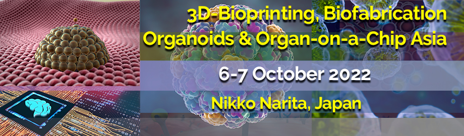
3D-Bioprinting, Biofabrication, Organoids & Organs-on-Chips Asia 2022
|
Lab-on-a-Chip and Microfluidics Asia 2022
| 
2022年10月6日(木) |
Please Refer to the Lab-on-a-Chip and Microfluidics Asia 2022 Track Agenda for Details of Programming on Thursday, 06 October 2022 |
| |
2022年10月7日(金)
08:00 | Morning Coffee, Tea and Networking in the Exhibit Hall | 09:00 | The Recent State of Oligonucleotide Therapeutics for Neurological Disorders
Hideki Mochizuki, Professor and Chairman, Osaka University Graduate School of Medicine, Japan
Oligonucleotide therapeutics have a wide range of potential applications in the clinic owing to their highly different modes of action such as antisense, ligands and protein inhibitors. In the field of neurology, ASOs that modulate splicing of SMN2 and Dystrophin pre-mRNA were approved for spinal muscular atrophy and Duchenne muscular dystrophy, respectively. siRNA encapsulated in lipid nanoparticle that degrades TTR mRNA was also available for hereditary ATTR amyloidosis. Many others are being developed worldwide, targeting neurological disorders caused by gene mutations and neurodegenerative diseases such as Huntington's disease, amyotrophic lateral sclerosis (ALS), Parkinson's disease (PD), multiple system atrophy (MSA) and Alzheimer's disease. Beyond pre-clinical trials, several clinical trials of oligonucleotide therapeutics are ongoing for neurological diseases, whereas phase III trials of tominersen (for Huntington's disease) and tofersen (for ALS) closed due to failing to attain their primary efficacy endpoints in 2021. We have been developing ASO suppressing SNCA expression as a potential therapy for MSA and PD. In this symposium, we will review the recent state of oligonucleotide therapeutics for neurological diseases including basic science, approved drugs and clinical trials. | 09:30 | Electrochemical Analysis of Vasculature-on-a-Chip and Vascularized-3D-Model-on-a-Chip
Yuji Nashimoto, Associate Professor, Institute of Biomaterials and Bioengineering, Tokyo Medical and Dental University, Japan
During this decade, bioengineering technologies to make a perfusable vascular model and integrate it with a three-dimensional culture model shows great advancement. However, analytical systems to evaluate vascular function and its effects are still limited. In this presentation, the electrochemical platform to evaluate vascular permeability, topography, and the transvascular flow effects on 3D tissue will be demonstrated. The analytical platform is promising for read-outs of the functionality of the vascular model and vascularized 3D model in a microphysiological system. | 10:00 | Microphysiological Systems (MPS) by Designing the Interface of Epithelial and Endothelial Cells
Ryuji Yokokawa, Professor, Department of Micro Engineering, Kyoto University, Japan
Microfluidic devices have become popular in many life science fields, including stem cell research. As a microfabrication scientist, I have been proposing new assay systems as microphysiological systems (MPS). The assay systems that mimic the functions of human biological organs can be constructed on a chip to measure physiological functions that are difficult to measure on a culture dish. We have employed two approaches to create the interface between organ cells and vascular networks in MPS: a two-dimensional method in which organ cells and vascular endothelial cells are co-cultured on the top and bottom surfaces of a porous membrane coated with an extracellular matrix, such as Transwell (2D-MPS), and a three-dimensional method in which the spontaneous patterning ability of vascular endothelial cells is utilized (3D-MPS). A 2D-MPS, renal proximal tubule model, evaluates albumin and glucose reabsorption and nephrotoxicity, while the glomerular filtration barrier model evaluates inulin and albumin filtration mechanisms. I will also present recent results on the development of a co-culture system of organoids and vascular network as a 3D-MPS. Kidney and brain organoids were cultured on a vascular network to demonstrate their maturation and vascularization. The on-chip vascular network is expected to expand from basic researches including vascular biology to evaluate the correlation between shear stress and vascular morphogenesis. | 10:30 | Mid-Morning Coffee, Tea and Networking in the Exhibit Hall | 11:00 |  | Keynote Presentation From Lab to Fork: 3D Tissue Engineering For Meat Production
Shoji Takeuchi, Professor, Center For International Research on Integrative Biomedical Systems (CIBiS), Institute of Industrial Science, The University of Tokyo, Japan
Research on "cultured meat," as typified by cultured hamburgers and chicken nuggets, has been studied world wide. These were made from randomly arranged muscle cells, so-called "minced meat." In contrast, our research group has been working on the in vitro fabrication of 3D structures of muscle tissue with the goal of realizing steak meat with its original texture. Bovine muscle tissue in the shape of a dice steak (1.0 cm x 0.8 cm x 0.7 cm) was prepared by forming a gel containing myoblasts grown from bovine muscle satellite cells into a sheet shape, stacking the sheet with both ends fixed to anchors, and culturing it. Myofibers in the tissue showed sarcomere-like structures stained with anti-a-actinin antibodies, suggesting that the myofibers were not just an aggregate of myoblasts, but that myoblasts fused with each other and underwent differentiation. In addition to these results, the latest developments will be presented in this talk. |
| 11:30 |  The Next Evolution in Microfluidic 3D Printing The Next Evolution in Microfluidic 3D Printing
Hemdeep Patel, President, Co-Founder, CADworks3D
Over the last decade, 3D printing has changed the way designs are created, evaluated and iterated in all industries and in every facet of life. Since 2016, CADworks3D has been an active player in the world of microfluidics & bio-engineering by showcasing how 3D printers can radically improve the cycle of design, evaluation and iteration. The CADworks3D line of microfluidic 3D printers and 3D materials has users to test a wide range of devices from clear microfluidic encapsulated chips to master molds used for casting PDMS devices. Our newest open source ProFluidics 285D microfluidic 3D printer equipped with updated optics, a next generation projector and advanced software will allow users to reproduce features that are more true to the design. Furthermore, the tell sign of pixel burn and shadow that was characteristic of the previous generation 3D printers are now a thing of the past allowing users to create smoother, superior quality surfaces finish. The ProFluidics 285D will allow users to create high quality clear microfluidic devices and master molds for casting PDMS devices.
| 12:00 | Light-Induced 3D Bioprinting Technologies
Daniel Nieto, Head of Biofabrication and Tissue Engineering unit. , University of Santiago de Compostela, Spain
An overview of some photo curing-based bioprinting technologies, including Digital Light Projection, Volumetric bioprinting and a light-based biopen for biomedical applications is presented. | 12:30 | Networking Lunch in the Exhibit Hall -- Visit Exhibitors and View Posters -- Japanese Bento Box Lunch | 14:00 | Biomimetic Vascular Constructs Using Three-Dimensionally (3D) Printed Porous Molds
Michinao Hashimoto, Associate Professor, Singapore University of Technology and Design, Singapore
We present a method to fabricate anatomically relevant vascular models using 3D-printed molds. Advanced biofabrication methods-sacrificial molding, direct ink writing, coaxial bioprinting, and embedded bioprinting-have enabled the fabrication of vascular models with intricate 3D architecture. Despite their advances, however, achieving full anatomical mimicry of native vasculature (such as freestanding, branching, multilayered, perfusable, and mechanically stretchable) remains to be challenging. In this work, we demonstrated an alternative biofabrication method for freestanding cell-laden vascular constructs with complex 3D architecture. The fabrication is achieved by employing a two-part mold consisting of porous hydrogels. The diffusion of calcium chloride (Ca2+) ions from the mold prompted dynamic crosslinking of the alginate-containing hydrogels in the radially inward direction to form a tubular construct. The same approach was extended to employing molds with complex shapes to achieve intricate 3D vascular architecture. The fabricated vascular models may be laden with smooth muscle cells (SMCs) and endothelial cells (ECs) in the multilayered arrangement. Lastly, vascular constructs with anatomically accurate geometries (e.g., constructions, bifurcation) and mechanical stress (e.g., cyclical motion) were readily fabricated. These vasculature models with increased biomimicry should benefit future research in mechanistic understanding of cardiovascular diseases and their therapeutic intervention. | 14:30 | 3D Modeling of Vascularized Barrier Tissues and Diseases For Preclinical Studies
Min Jae Song , Staff Scientist, National Center for Advancing Translational Sciences (NCATS), United States of America
In vitro three dimensional (3D) cellular models enable the study of multicellular interactions within functional tissue microenvironments. The enhanced physiological relevance of these complex 3D cellular models has opened the possibility of developing human-pathologically relevant disease assays for preclinical drug discovery and development studies. However, the increased cellular and structural complexity of these 3D cellular assays pose a significant technical challenge for their morphological and physiological validation, and use for pharmacological testing. Using 3D bioprinting techniques, we have established a robust and versatile method to engineer human vascularized tissues in a multiwell format. The bioprinting-based approach, used to biofabricate vascularized tissues, included a biodegradable polymer scaffold that enabled the addition of epithelia, in a transwell format. Several human barrier tissue models with vascularization were produced, including skin, peritoneal, and ocular tissues. Once 3D models of “healthy” tissues were biofabricated and validated, disease tissue models were developed by introducing disease-relevant chemical inducers or diseased cells, like cancer cells, into the “healthy” tissues. Treatments of the disease models with FDA approved drugs or drugs in clinical trials were able to correct the disease phenotypes. The structural, functional, and pharmacological validation of these tissues is critical to enable the use of these 3D models to accelerate the drug development process by providing pre-clinical data that it is more predictive of clinical outcomes.
| 15:00 | Research and Development of Microphysiological Systems in Japan Supported by the AMED-MPS Project
Seiichi Ishida, Guest Researcher, National Institute of Health Sciences, Professor, Sojo University, Japan
Microphysiological Systems (MPS) is expected to be novel humanized in vitro test methods that addresses the unmet needs for new drug development. In Japan, the AMED-MPS project is being promoted under the leadership of AMED (the Japan Agency for Medical Research and Development) and Ministry of Economy, Trade and Industry(1). In this project, along with the development of Japan-original MPS for commercialization, there have been discussions on the technical requirements that need to be solved for MPS to be implemented in industry. Stakeholders from academia developers, suppliers of MPS, users of pharmaceutical industry, and the regulatory section are participating in these discussions actively. In this presentation, firstly the development status of Japan-original MPS will be introduced. And, secondly, we would like to present and discuss the concept of "technical requirements for industrial implementation of MPS" from the viewpoint of regulatory science based on the discussions in AMED-MPS project by showing how to establish “Context of Use” from unmet needs that MPS is required to solve and how to promote standardization of MPS. (1) Seiichi Ishida: Research and Development of Microphysiological Systems in Japan Supported by the AMED-MPS Project. Front. Toxicol. doi: 10.3389/ftox.2021.657765 | 15:30 | Mid-Afternoon Coffee and Tea Break and Networking in the Exhibit Hall | 16:00 |  | Keynote Presentation Bioprinting of Soft Tissues: Interplay Between Design and Function
Wai Yee Yeong, Professor, School of Mechanical & Aerospace Engineering, Nanyang Technological University, Singapore
Bioprinting technologies are enabling tools to achieve functional design of tissues and organs. While the technology is still at demonstration stage, there are a lot of potentials to utilize the physical processes of bioprinting to influence the functional properties of the printing tissues. In this talk, we will discuss about the interplay between design and functions of soft tissues enabled by bioprinting. Examples of extrusion and droplet-based bioprinting will be presented to illustrate different strategies of biomimetic design.
|
| 16:30 | Digital Light Processing (DLP) 3D Printing of Biological Tissues Using PEGDMA-based Bioinks
Shu-Yung Nina Chang, Research Fellow, Singapore University of Technology and Design, Singapore
3D bioprinting is an emerging approach to create biomimetic structures as it allows precise spatial control of the resulting tissue construct. In this study, we present the application of PEGDMA-based bioink to create complex biological tissues using a DLP printer. | 17:00 | Biofabrication of Three-Dimensional (3D) Cell-Laden Constructs by Thermoresponsive Release of Photo-Patterned Hydrogels
Xiaolei Nie, Research Fellow, Singapore University of Technology and Design, Singapore
We report on a unique method of biofabrication to pattern cell-laden grafts on a thermoresponsive surface for controlled release and incubation. The assembly of the released grafts into 3D structures was also demonstrated. | 17:30 | Microfluidics-based Generation of Curcumin-loaded Microfibers for Biofilm Remedy Using Photodynamic Therapy
Kajal Sharma, PhD Student, IITB-Monash Research Academy, India
The rapid increase in multidrug-resistant biofilm infections is a major concern for global health. Thus, highly effective therapy is required for the treatment of biofilm-related infections. In this study, curcumin loaded alginate microfibers were generated using the microfluidic technique. In this strategy, the alginate microfiber is used as a carrier for the encapsulation of the curcumin, which then is irradiated with blue light, photodynamic therapy (PDT) to assess the efficacy of combined therapy against the drug-resistant S. aureus. The advantage of utilizing PDT is the non-antibiotic mode of bacterial cell inactivation. In the presence of blue light, PDT the curcumin loaded alginate microfiber has shown a good eradication activity against the infections caused by multi-drug resistant S. aureus. We generated different diameters of the curcumin loaded alginate microfiber by varying the system flow rates, and the process was imaged using an inverted optical microscope (Nikon Eclipse Ti-S, Japan) connected to a high-speed camera (FASTCAM Mini UX100, Japan). The curcumin loaded microfiber was characterized using Scanning electron microscopy, Fourier Transform Infrared spectroscopy (FTIR), and X-ray diffraction (XRD). Further, the efficacy of curcumin loaded alginate microfibers in presence of blue light has been evaluated against biofilm forming bacterial S. aureus (NCIM 5718) by confocal and optical microscopy. In summary, in this work, we present an efficacious and cost-effective micro-fibrous scaffold for the controlled release of curcumin to treat biofilm in presence of blue light. | 18:00 |  | Keynote Presentation 3D Bioprinting: From Organ Model to Tissue Repair
Yong He, Professor, Zhejiang University, China
In this talk, we reported the recent progress in 3D bioprinting of our group. 1) Standardizing the bioinks and a framework is given for the analysis of printability during projection based 3D bioprinting(PBP); 2) How to directly print cell-laden structures with effectively vascularized nutrient delivery channels? 3) How to mimic the complex extracellular matrix with near field direct writing? |
| 18:30 | Close of Conference |
|